Blogerroom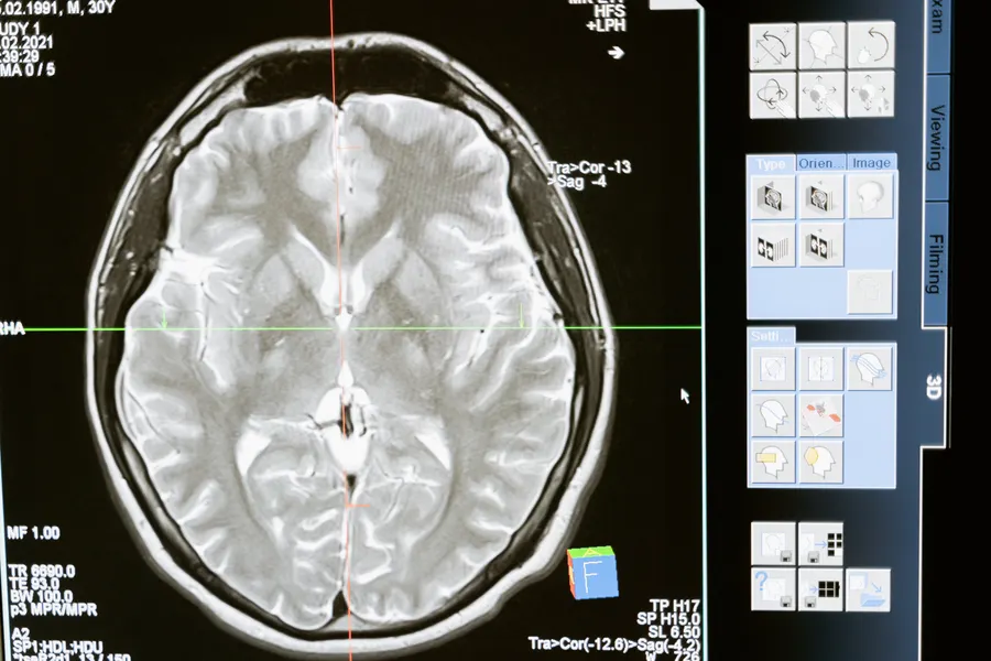
UCLA Reveals Hidden Rules That Build the Human Brain
Two UCLA studies found brain stem cells shift what neurons they make based on glucose metabolism and early thalamic contact.
A stem cell making billions of choices before you're even born
Before a baby takes its first breath, its brain has already been assembled through an almost incomprehensible number of cellular decisions. At the center of that process sits a single, remarkable type of stem cell called radial glia, which generates much of the cerebral cortex, the seat of thought, memory, and language, and is thought to drive much of what makes the human cortex larger and more complex than that of any other species. Two companion studies from UCLA, published simultaneously this week in Cell and Science, identify two previously unrecognized forces shaping the choices these cells make: how they metabolize glucose, and physical contact from a neighboring brain structure that arrives far earlier than anyone expected.
Together, the two papers, both from the lab of Aparna Bhaduri at UCLA, reframe radial glia's surroundings, its nutrients and its physical connections, as active, decision-shaping influences on brain development, rather than a passive backdrop the cells simply operate within.
The metabolism study: sugar as an instruction, not just fuel
The first study focused on how radial glia process glucose, and the finding runs against a common assumption in cell biology: that metabolism is mostly a background process, quietly powering whatever a cell has already been instructed to do by its genetic program. Bhaduri's team found that radial glia rely heavily on a specific metabolic route called the pentose phosphate pathway, which uses glucose to generate building blocks needed by rapidly dividing cells. When researchers reduced glucose availability or directly disrupted that pathway, the stem cells didn't just slow down or malfunction generically. They changed what they actually produced, shifting toward generating more inhibitory neurons and other cell types that normally appear later in cortical development.
"What was surprising is that metabolism isn't just a passive thing that happens in the background," Bhaduri said. That's a meaningful reconceptualization. If glucose metabolism itself can push a stem cell toward producing different neuron types altogether, rather than simply providing more or less energy for a predetermined developmental program, then nutrient availability during pregnancy becomes a genuinely active variable in how the brain gets built, not just a resource that needs to be sufficient in the background.
The thalamus study: a signal that arrives suspiciously early
The second study, led by first author Claudia Nguyen, tackled a different and longstanding puzzle. Anatomists have long known that thalamic projections, the long, wire-like fibers extending from the thalamus, a deep brain structure that relays sensory and other information throughout the nervous system, reach the developing cortex much earlier than when their final, functional connections actually form. That timing gap raised an obvious question nobody had definitively answered: what are these projections doing there so early, well before they're needed for their eventual signaling role?
Using human stem cell-derived brain "assembloids," fused three-dimensional structures combining thalamic and cortical tissue grown from stem cells, the UCLA team found a concrete answer. Thalamic projections make direct physical contact with radial glia during development, and that contact actively drives the stem cells to produce more of the specific neuron types most expanded in the human brain compared with other species, upper-layer neurons associated with the complex cortical circuitry that distinguishes human cognition. In other words, the thalamus isn't just waiting around for the cortex to finish developing before plugging in. It's actively participating in building the cortex it will eventually communicate with.
Why assembloids made this discovery possible
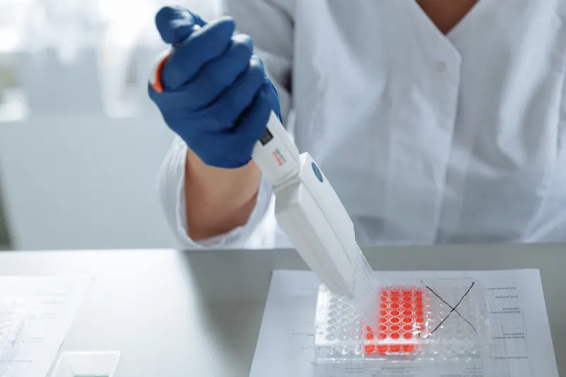
The methodology behind the thalamus finding deserves attention on its own. Studying live human fetal brain tissue at this level of experimental precision, deliberately manipulating physical contact between specific cell populations, isn't ethically or practically feasible. Brain assembloids solve that problem by fusing separately-grown organoids representing different brain regions, in this case cortical and thalamic tissue, into a single connected structure, letting researchers observe and manipulate genuine cross-region interactions that would otherwise be impossible to study directly in a developing human embryo.
That technique is part of why both of these findings arrived now rather than years ago. Assembloid and organoid models have matured considerably as experimental tools over the past several years, giving researchers access to questions about human-specific brain development that were previously answerable only through animal models poorly suited to capturing what actually makes the human cortex distinctly human in the first place.
What goes wrong when these signals get disrupted
Both studies point toward the same broader stake: understanding what happens when these newly identified influences, metabolic and physical, go awry. Radial glia largely disappear before birth once their neuron-generating work is done, but cells resembling them can reemerge later in life for reasons that remain unclear, specifically in certain brain cancers. A stem cell population this central to normal development, and this capable of reappearing abnormally in disease, makes understanding its full range of regulatory signals a genuinely high-stakes research question, not just an academic curiosity about fetal development.
The metabolic findings carry a particularly direct clinical resonance. If glucose processing genuinely shapes which neurons radial glia produce, that raises real questions about how maternal nutrition, metabolic conditions during pregnancy, or disruptions to glucose regulation might influence fetal brain development in ways researchers hadn't previously connected to cortical cell-type diversity. Similarly, if thalamic contact actively shapes upper-layer neuron production, disruptions to that specific signaling pathway, whether from genetic factors or developmental timing issues, become a plausible mechanistic thread connecting early brain wiring problems to neurodevelopmental disorders that show up much later in a child's life.
What these findings don't yet establish
It's worth being precise about the limits here. Both studies were conducted using organoid and assembloid models, laboratory-grown structures that capture important features of early human brain development but remain simplified representations, not full replicas of an actual developing fetal brain complete with vasculature, immune signaling, and the full three-dimensional architecture of gestation. Translating these mechanistic findings into direct clinical guidance, about maternal nutrition during pregnancy, for instance, would require considerably more research connecting these cellular-level metabolic effects to actual outcomes in real pregnancies, a gap the current studies don't attempt to close.
What they do establish, convincingly, is that radial glia's environment, the sugar available to it and the physical company it keeps, actively participates in deciding what kind of brain gets built. That's a genuinely new layer to add to a process scientists have been mapping in ever-finer detail for decades, and it opens two concrete new research directions, metabolic regulation and cross-region physical signaling, that weren't clearly on the map before this week.
Written by
Dr. Anand Sharma
Doctor and science communicator.